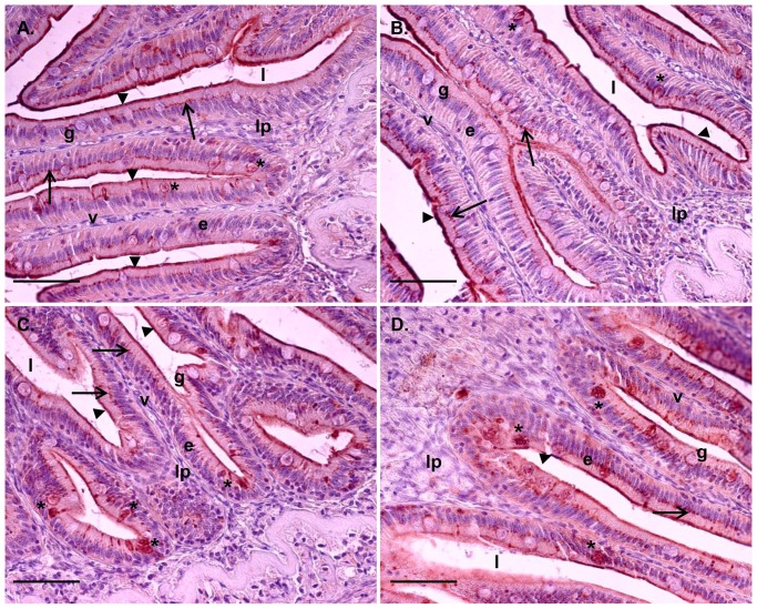
Figure 3. Tissue localization of Mx immune staining in the proximal and distal intestine.
Tissue sections, after 6 days of cohabitant challenge, from proximal intestine (A and B) and distal intestine (C and D). Scale bar 50 µm. Epithelial cells (e) lining the villi (v) show supra nuclear presence of Mx (arrow). Mx is also present in the most apical part of the enterocytes in the micro villi region (arrowhead). Staining could also be found in goblet cells (g) (*). The lamina propria (lp) and intestinal lumen (l) are featured for clarity.

