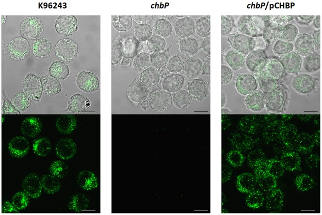
Figure 2. Confocal micrographs of CHBP expression and localization in U937 cells infected with B. pseudomallei.
PMA-activated U937 cells were separately infected with three strains of B. pseudomallei (K96243, chbP mutant or chbP/pCHBP strain). After 6 h, infected cells were fixed, permeabilized andstained using purified rabbit anti-CHBP antibody detected with anti-rabbit Ig-Alexa Fluor488 (Molecular Probes). The bottom panel shows the localization of CHBP and the top panel merges this signal with differential interference contrast (DIC) images showing the position of infected cells. Scale bars, 20 µm.

