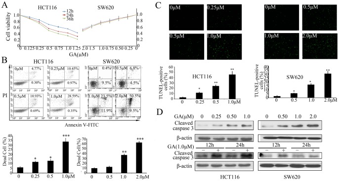
Figure 1. GA promotes apoptosis in colorectal cancer cells.
(A) HCT116 and SW620 cells were treated with increasing concentrations of GA for 12 h, 24 h or 36 h, and the cell viability index was measured by MTT assay. (B) HCT116 and SW620 cells were treated with increasing concentrations of GA for 24 h, cell apoptosis was detected by annexin-V fluorescein isothiocyanate (FITC) and propidium iodide (PI) double staining followed by flow cytometry analysis. Dot plot display of Annexin-V FITC-fluorescence versus propidium iodide fluorescence is shown in logarithmic scale. Living cells tested negative for both annexin V-FITC and PI. Populations testing annexin V positive/PI negative were classified as early-stage apoptotic cells, and double-positive cells were classified as dead cells. Bar diagram showing the percentage of dead cells after different treatments. (C) HCT116 and SW620 cells were treated with increasing concentrations of GA for 24 h, dead cells were detected by TUNEL assay. The TUNEL-positive cells were counted from at least 100 random fields. (D) Immunoblot analysis of cleaved-caspase 3 from lysates of HCT116 and SW620 cells treated with various concentrations of GA for 24 h, or treated with 1 µM GA for 12 h and 24 h.

