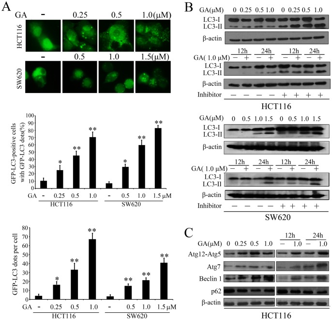
Figure 3. GA initiates autophagy in colorectal cancer cells.
(A) HCT116 and SW620 cells transfected with a pEGFP-LC3 plasmid were treated with indicated concentrations of GA for 24 h. Cells were defined as positive if they had 5 or more GFP-LC3 dots in the cytoplasm. The percentage of the cells with GFP-LC3 dots and the average number of GFP-LC3 dots per cell were analyzed from at least 100 random fields. (B) Immunoblot analysis of the conversion of LC3-I to LC3-II in HCT116 cells and SW620 cells after treated with indicated concentrations of GA for 24 h, or with 1.0 µM of GA for 12 h and 24 h in the absence or presence of lysosomal inhibitors (E64d and pepstatin each at 10 µg/ml). (C) Immunoblot analysis of the expression level of Atg12-Atg5 conjugate, Atg7, Beclin 1 and p62 after treated with indicated concentrations of GA for 24 h, or with 1 µM of GA for 12 h and 24 h. Actin served as a loading control. * p<0.05; ** p<0.01.

