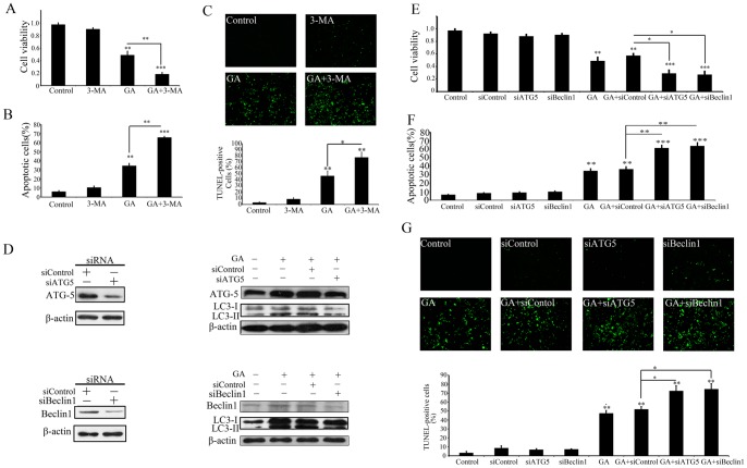
Figure 4. Blockage of autophagy enhances GA-induced apoptosis.
(A–C) HCT116 cells were treated with vehicle control (1‰ DMSO, Control), 3-MA, 1 µM GA (GA), or 1 µM GA in the presence 3-MA (GA+3-MA) for 24 h. And then the cell viability was evaluated by MTT assay (A), and the apoptotic effect was detected by PI/annexin-V staining (B) and TUNEL assays(C). (D) Immunoblot detection of the expression of ATG5, beclin-1 and LC3 in HCT116 cells treated with GA in the present or absent with siATG5 or siBeclin 1. (E–G) HCT116 cells were treated with Lipofectamine 2000 (Control), control siRNA (siControl), siATG5 (siATG5), 1 µM GA (GA), GA in the presence control siRNA (GA+siControl), siATG5 (GA+siATG5) or siBeclin1 (GA+siBeclin 1) for 24 h. And then the cell viability was evaluated by MTT assay (E), and the apoptotic effect was detected by PI/annexin-V staining (F) and TUNEL assays (G). * p<0.05; ** p<0.01.

