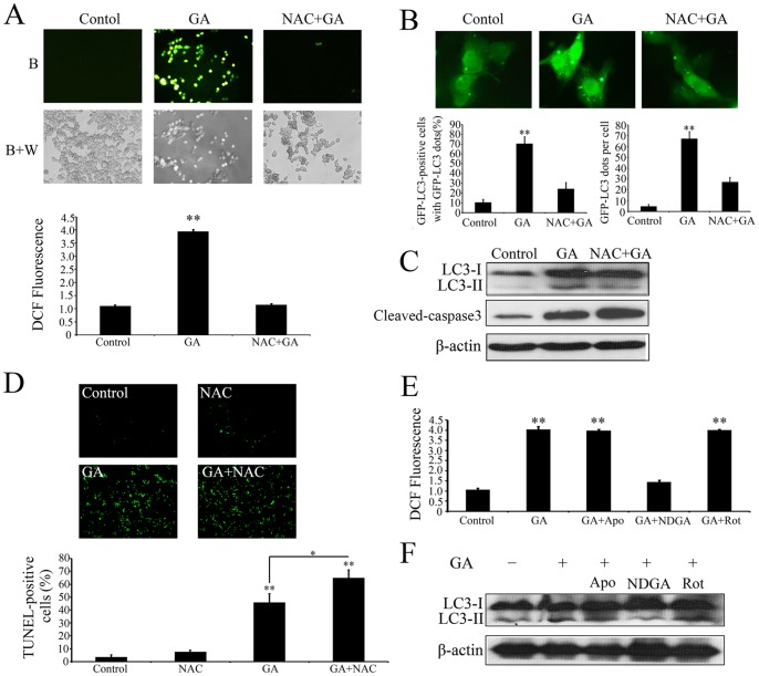
Figure 6. ROS is required for GA-induced autophagy.
(A) Intracellular ROS in HCT116 cells treated with 1‰ DMSO (Control), 1.0 µM GA (GA), or 1.0 µM GA in the presence NAC (10 mM) (GA+NAC) for 24 h were detected by staining cells with 2′,7′-dichlorofluorescein diacetate under blue (B), or blue and white excitation (B+A). The DCFH-DA signal was measured using a Molecular Devices SPECTRAMAX M5 fluorimeter. (B) HCT116 cells transfected with a pEGFP-LC3 plasmid with or without GA (1.0 µM) or/and NAC (10 mM) for 24 h. Cells were defined as positive if they had 5 or more GFP-LC3 dots in the cytoplasm. The percentage of the cells with GFP-LC3 dots and the average number of GFP-LC3 dots per cell were analyzed from at least 100 random fields. ** p<0.01. (C) Immunoblot analysis of the conversion of LC3-I to LC3-II and expression of cleaved-caspase 3. (D) HCT116 cells treated with or without GA (1.0 µM) or/and NAC (10 mM) for 24 h, cell apoptosis was detected by TUNEL assay. The TUNEL-positive cells were counted from at least 100 random fields. (E) HCT116 cells were treated with or without GA or/and the antioxidants Apocynin, rotenone and nordihydroguaiaretic acid (NDGA) for 24 h, and then the intracellular ROS were measured using a Molecular Devices SPECTRAMAX M5 fluorimeter. (F) HCT116 cells were treated with or without GA or/and the antioxidants Apocynin, rotenone and nordihydroguaiaretic acid (NDGA) for 24 h, and then the conversion of LC3-I to LC3-II were detected by immunoblot analysis.

