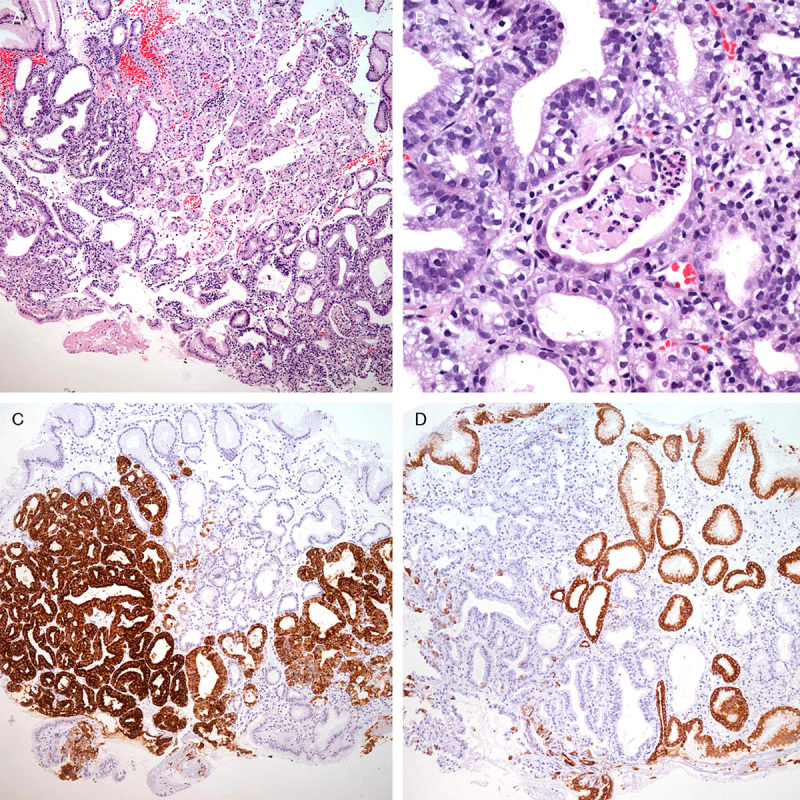
FIGURE 1.

Initial endoscopy findings of patient #1. A, The lesion was composed of packed complex pyloric-type glands lined by cuboidal or columnar cells. B, The tumor cells showed enlarged rounded nuclei and loss of polarity with pale to eosinophilic cytoplasm. C, Most glands were strongly positive for MUC6 protein. D, The superficial layer was positive for MUC5AC.
