
FIGURE 2.
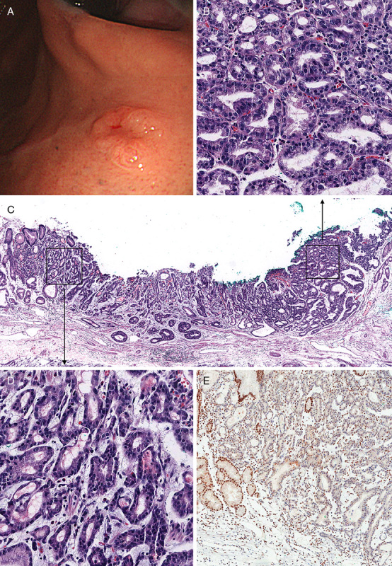
A, Endoscopic findings at the 2-year follow-up for patient #1. A, Dome-like polypoid lesion measuring 1.0×0.6 cm with central ulceration was noted. B, Pathologic examination of endoscopic submucosal dissection specimen showed PGA with round nuclei and amphophilic cytoplasm, which is similar to that of the original biopsy. C, Direct transformation of PGA to invasive adenocarcinoma. D, Adenocarcinoma with invasion of glands into lamina propria. E, Loss of expression of MLH1 in adenocarcinoma and PGA.