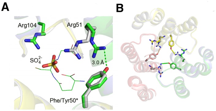
Figure 5. Positional shift of Arg51 and loss of salt bridge at the active site of mLTC4S.

A. Close up of the mLTC4S complex with SO4 2−, showing a shift in the position of Arg51 due to Phe50Tyr exchange. Human LTC4S is colored in green and mLTC4S is colored in gray. GSH is shown as green “lines”. *indicates that it is positioned on the neighboring subunit. B. Trimer of mLTC4S showing the amino acid exchange at position 50 where Phe in mLTC4S fails to make a salt bridge with Arg51. In hLTC4S, the Tyr50-Arg51 couple will likely contribute to trimer stability.
