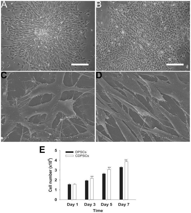
Figure 1. Morphology and proliferative potential of DPSCs and CDPSCs.
Both DPSCs (A) and CDPSCs (B) isolated from dental pulp were spindle-shaped and fibroblast-like under the light microscope. Both DPSCs (C) and CDPSCs (D) have a fibroblast-like appearance with long cytoplasmic processes and many filopodia using the scanning electron microscope. (E) CDPSCs have a higher proliferative potential compared with DPSCs (**p<0.01). Scale bar = 100 µm. Each experiment was repeated in triplicate.

