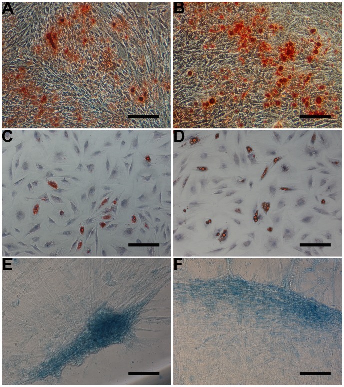
Figure 2. Multilineage differentiation potential of DPSCs and CDPSCs.
Mineralization assay in DPSCs and CDPSCs. Mineralized nodules formed by DPSCs (A) and CDPSCs (B) were detected by alizarin red S staining after 3 weeks of culture in mineralized-induced media. Adipogenic differentiation, visualized by oil red O staining, showed lipid vacuoles in DPSCs (C) and CDPSCs (D). Chondrogenic differentiation was visualized by alcian blue staining of DPSCs (E) and CDPSCs (F), demonstrated by the accretion of sulfated matrix. Scale bar = 100 µm. Each experiment was repeated 3 times.

