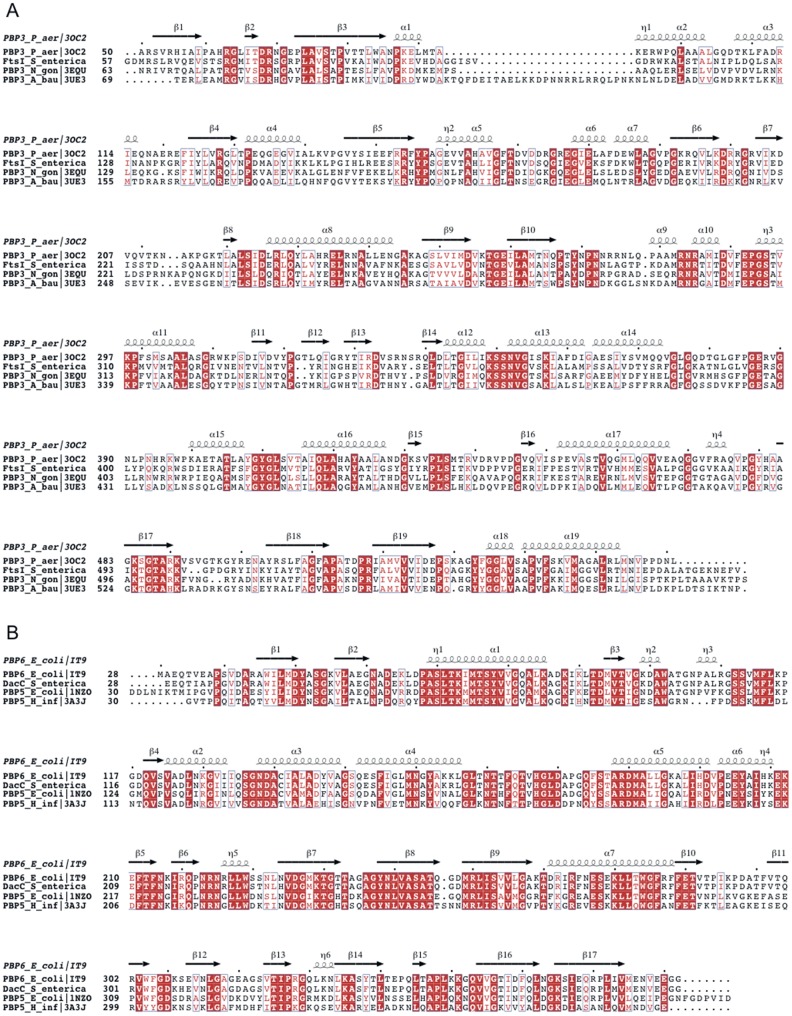
Figure 1. Sequence alignments of S. enterica FtsI (A) and DacC (B) with PBPs with known structure.
Secondary structure is indicated above the alignment. Strictly conserved residues are shown in white on red background, residues with conserved physico-chemical properties are shown in red on white background. Mutation sites in this study are marked with asterisks below the alignment. The figure was prepared using ESPRIPT [34].

