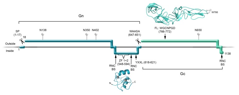
Figure 3.
Schematic Representation of Hantavirus Glycoprotein Processing and Functions. The Gn (dark cyan) and Gc (light cyan) glycoprotein ectodomains, transmembrane regions and endodomains are represented according to their location relative to the membrane (horizontal grey line). The Signal peptide (SP) and WAASA sequences indicate the cleavage sites within GPC. N represents the location of asparagine residues that are likely to carry glycosylations; the numbers following these letters indicate the corresponding residue number within GPC of ANDV. ZF 1 + 2 indicates the location of the two zinc finger domains, RNC-BS indicates the suggested ribonucleocapsid binding sites. The motif YxxL represents residues involved in ubiquitination. FL indicates the location of the putative fusion loop of ANDV Gc. The molecular structure of the ANDV Gn-CT zinc finger domains (dark cyan) was obtained from the NCBI databank, PDBid: 2K9H [24]. The molecular model for the ANDV Gc fusion protein ectodomain was developed previously [29]. All ribbon diagramsof molecular structures were rendered in the PyMOL Molecular Graphics System [61].

