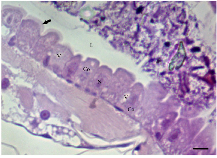
Figure 1.
Photomicrograph of the midgut of Aedes aegypti third-instar larvae (Diptera: Culicidae) stained with hematoxyline and eosin (HE). Columnar cells (Co), central nucleus (N), some cytoplasm vacuoles (v) and pink brush border (arrow) in the apical surface of the cells in larvae of controls group. L = lumen. Bar: 20 μm.

