
Figure 3.
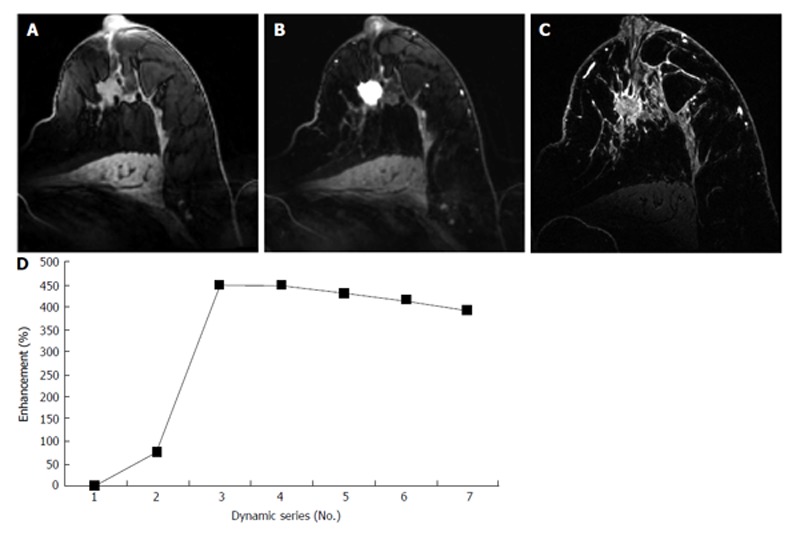
A 62-year-old patient with nipple withdrawal, finally diagnosed as ductolobular carcinoma. A, B and C: Axial T1-weighted gradient-echo images obtained at 7 T before and after contrast injection. An irregular mass with spiculated margins can be observed on pré-contrast imaging (A). An intense homogeneous enhancement (B) and a rapid wash-out kinetic curve (D) can be observed following contrast administration. In Figure 3C, an ultra-high-resolution T1-weighted gradient-echo sequence with fat suppression was performed, and the morphological aspects of the lesion can be more clearly seen.