Figure 3.

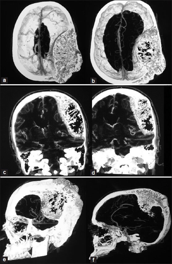
Horizontal, coronal and sagittal angio-enhancing, standard, and 3D sections showing mild vascularity of the lesion and its interface with the dura mater

Horizontal, coronal and sagittal angio-enhancing, standard, and 3D sections showing mild vascularity of the lesion and its interface with the dura mater