


Figure 4.
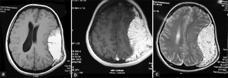
Axial T1W and T2W MR images showing hyperintense trabeculated lesion expanding the calvarial bone both intra- and extra-cranially. The tumoral tissue extended within the diploe of the adjacent frontal, temporal, occipital bones, and toward the midline suture