


Figure 7.
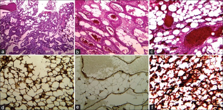
a-c, H and E staining of the specimen with different magnifications, showing mature adipose tissue with a prominent vascular component. The lipomatous components were surrounded by bands of fibrous connective sheaths containing foamy macrophages and scattered mast cells ‘chicken wire appearance’. Bone spicules remained within the lesion contained osteocytes but no osteoclast and osteoblast cells. The vascular components consisted of thin-walled vessels of different sizes and small groups of capillaries containing erythrocytes and thrombus formations. d and e, The IHC staining using antibodies for CD34 and S100 protein showing the endothelial vascular sheaths and foamy lipomatous compartments. d: Sudan III staining without mounting, demonstrating neutral lipids component in the tissue