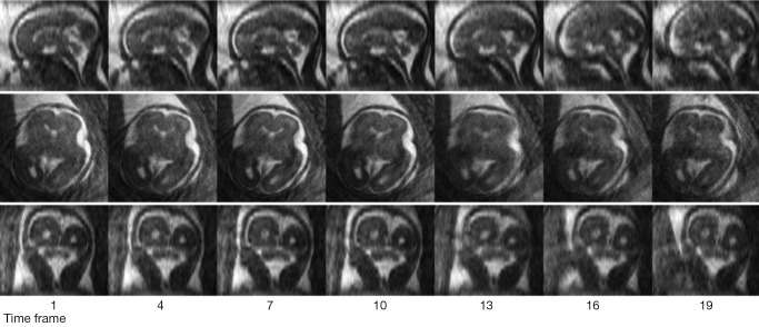
Figure 2.
Time-resolved 3D fetal images were acquired during a 3-minute scan. A total of 20 frames were reconstructed, with a temporal resolution of 9 s. Seven representative frames at every four frames are shown in the axial, sagittal and coronal planes. It can be observed that the brain has turned slightly towards the midline over the course of the scan.

