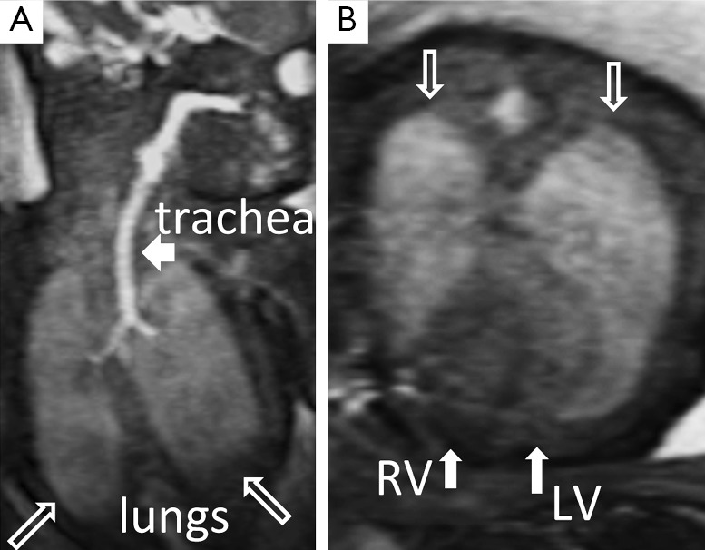
Figure 4.

(A) Coronal view of the fetal lungs (hollow arrows) and trachea (short arrow); (B) Axial view of the fetal lungs (hollow arrows) and long-axis view of the non-ECG-gated fetal heart (solid arrows). LV, left ventricle; RV, denotes right ventricle.
