
Figure 5.
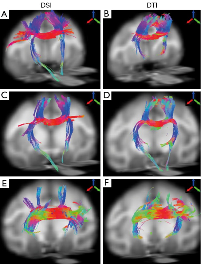
Illustration of fiber tractography of an adult rhesus monkey brain using DSI (left) and DTI (right). From top to bottom: DTI- and DSI-derived fibers across posterior midbody (A,B), isthmus (C,D) and splenium (E,F), respectively. Arrows indicate the color-coding of spatial orientations. DSI, diffusion spectrum imaging; DTI, diffusion tensor imaging.