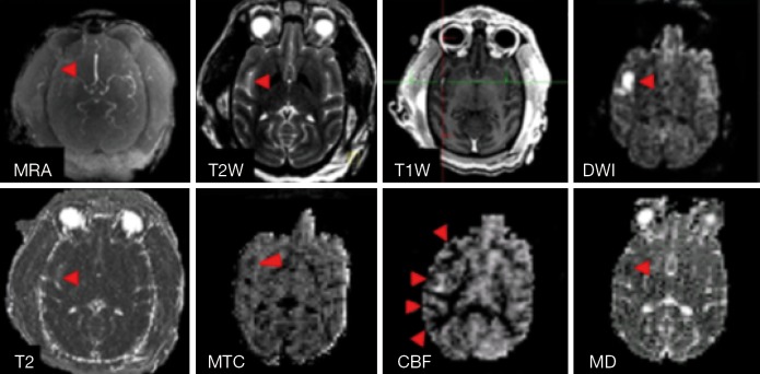
Figure 3.
Illustration of stroke lesion. Top, MR Angiography, T2W, T1W, and DWI images of an adult stroke monkey (RLB6) post reperfusion; bottom: quantitative MRI measures (T2, MTC, CBF, and MD maps) of the same monkey. The monkey (RLB6) was induced with 3-hour transient MCA occlusion. Arrows: stroke-injured regions. MRA, MR angiography; T2W, T2-weighted imaging; T1W, T1-weighted imaging; MTC, magnetization transfer contrast; CBF, cerebral blood flow; MD, mean diffusivity; MCA, middle cerebral artery.

