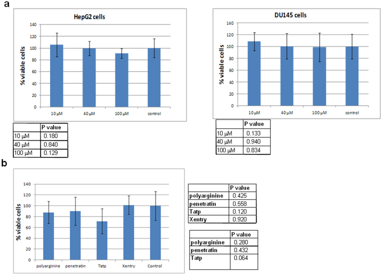
Figure 8. Xentry is non-toxic to HepG2 and DU145 cells.
(a) HepG2 and DU145 cells were incubated for 24 h with 0, 10, 40, and 100 μM of FITC-labelled D-isomeric Xentry (lclrpvg) for 24 h, and cell viability measured using the WST-1 assay. The mean percentage of viable cells was plotted ± SD. The statistical significance of the differences in cell viability compared to cells that were not treated with Xentry is shown in the tables beneath each graph. (b) HepG2 cells were incubated for 24 h with biotin- labelled Xentry (LCLRPVGGGRRRQQQQQQRRR), penetratin (GRKKRRQRRRPPQGGRRRQQQQQQRRR), polyarginine (RRRRRRRRRQQQQQQRRR) and Tatp (RQIKIWFQNRRMKWKKGGRRRQQQQQQRRR) peptides at final concentrations of 10 μM. Cell viability was measured using the WST-1 assay. The mean percentage of viable cells was plotted ± SD. Untreated HepG2 cells were included as controls. The statistical significance of the differences in cell viability after treatment with each of the CPPs compared to untreated control cells is shown in the top right-hand table. The statistical significance of the differences in cell viability after treatment with Xentry compared to the other CPPs is shown in the bottom right-hand table.

