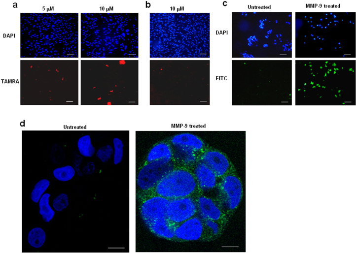
Figure 9. Linear multivalent and heparin mimic-conjugated forms of Xentry are not cell-penetrating, but have utility as protease-activatable peptides.
A TAMRA-labelled divalent D-isomer (lclrpvggggggggggggggglclrpvg) (a), and a divalent L-isomer (LCLRPVGGGGGGGGGGGGGGGLCLRPVG) (b) of Xentry are unable to penetrate HepG2 cells. The peptides were incubated for 3 h with HepG2 cells at concentrations of 5 and/or 10 μM, as indicated. Cells were fixed and cell fluorescence recorded using a Nikon E600 fluorescence microscope. Cell nuclei were stained blue with DAPI. Images were taken at 20× magnification. Scale bar represents 50 μm. The small amount of red fluorescence is not cell-associated. (c, d) Activatable forms of Xentry. (c) The ACPP lclrpvGGGGPLGLAGGlclrpvgk-FITC peptide at a final concentration of 10 μM was left untreated or incubated for 3 h with 0.2 μg of activated MMP-9, and then incubated with MCF-7 cells for 3 h. The cells were fixed and fluorescence was visualized and recorded using a Nikon E600 fluorescence microscope. Cell nuclei were stained blue with DAPI. Images were taken at 20× magnification. Scale bar represents 50 μm. (d) The ACPP GSY(sulfated)DY(sulfated)GGGGPLGLAGGlclrpvgk-FITC peptide at a final concentration of 10 μM was left untreated or incubated for 3 h with 0.2 μg of activated MMP-9, and then incubated with MCF-7 cells for 3 h. The cells were fixed and fluorescence was visualized and recorded using a Zeiss LSM 710 inverted confocal microscope. Cell nuclei were stained blue with DAPI. Images were taken at 63× magnification. Scale bar represents 10 μm.

