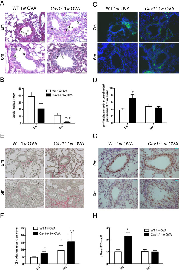
Figure 5.
Alterations of the airway remodeling features in mice after OVA challenge in Cav1-/- mice. Representative pictures of 1 week-OVA challenged WT and Cav1-/- mouse lung sections. (A,B.) Goblet cells metaplasia was detected using Periodic Acid Schiff staining. Positive cells (indicated by arrows) were counted in airways of 100-200 μm in an average of 5 airways per animals is reported (Bar graph). (C,D.) Detection of α-SMA positive cells by immunohistochemistry and reporting the surface of positive staining relative to the length of basement membrane (Bar graph). (E,F.) Detection of collagen depositon around the airways using Picrosirius red staining and quantification of the relative amount of collagen deposition (Bar graph). (G,H.) Detection of pSmad2 using immunohistochemistry. Activation of the canonical TGF–β signaling pathway was also determined by western blot. Densitometric quantitations were carried out using the whole lung homogenates of WT and Cav1-/- mice after a 1-week OVA challenge. Values are represented as relative protein levels of pSmad2 to total Smad2 (Bar graph). For all the measurement, * < p 0.05 represents significance by 2-way ANOVA between WT and Cav1-/- mice for the same age group, and # < p 0.05 represents significance by 2-way ANOVA between same age group for WT and Cav1-/- mice.

