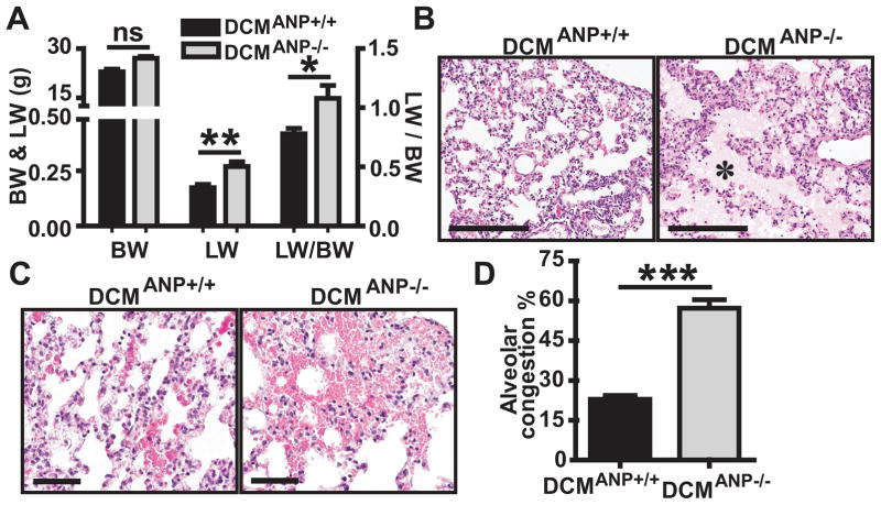
Figure 2. Enhanced heart dysfunction and pulmonary congestion in DCMANP−/− mice.
A. Lung wet weight (LW), body weight (BW) and LW:BW ratio (n = 12–23 in each group). B&C. Histological evaluation of lung congestion by H&E stain. B. DCMANP+/+ mice have mild interstitial edema while DCMANP−/− mice show severe interstitial and intra-alveolar edema (pink area, asterisks); C. DCMANP−/− mice have more punctate hemorrhagic lesions in alveolar capillaries than DCMANP+/+ mice. Scale bars=200μm. D. Quantitative analysis of total alveolar congestion area per 20× field. Results are means of averages of 12 randomly selected fields from 3 to 4 mice in each group. ***p<0.001, **p<0.01, *p<0.05, nsp>0.05.

