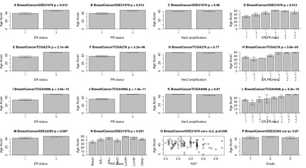
Figure 8.

Age acceleration in breast cancer. Panels in the first column (A,E,I,M) show that estrogen receptor (ER)-positive breast cancer samples have increased age acceleration in four independent data sets. Panels in the second column (B,F,J) show the same result for progesterone receptor (PR)-positive cancers. Panels in the third column (C,G,K) show that HER2/neu amplification is not associated with age acceleration. Panels in the fourth column (D,H,L) show how combinations of these genomic aberrations affect age acceleration. (N) Age acceleration across the following breast cancer types: Basal-like, HER2-type, luminal A, luminal B, and healthy (normal) breast tissue. (O) Ki-67 expression versus age acceleration. (P) Tumor grade is not significantly related to age accelerations, reflecting results from Additional file 14. Vertical grey numbers on the x-axis report sample sizes. The figure titles report the data source (GSE identifier from Gene Expression Omnibus or TCGA), and the Kruskal Wallis test P-value (except for panels (O,P), which report correlation test P-values). Error bars represent 1 standard error.
