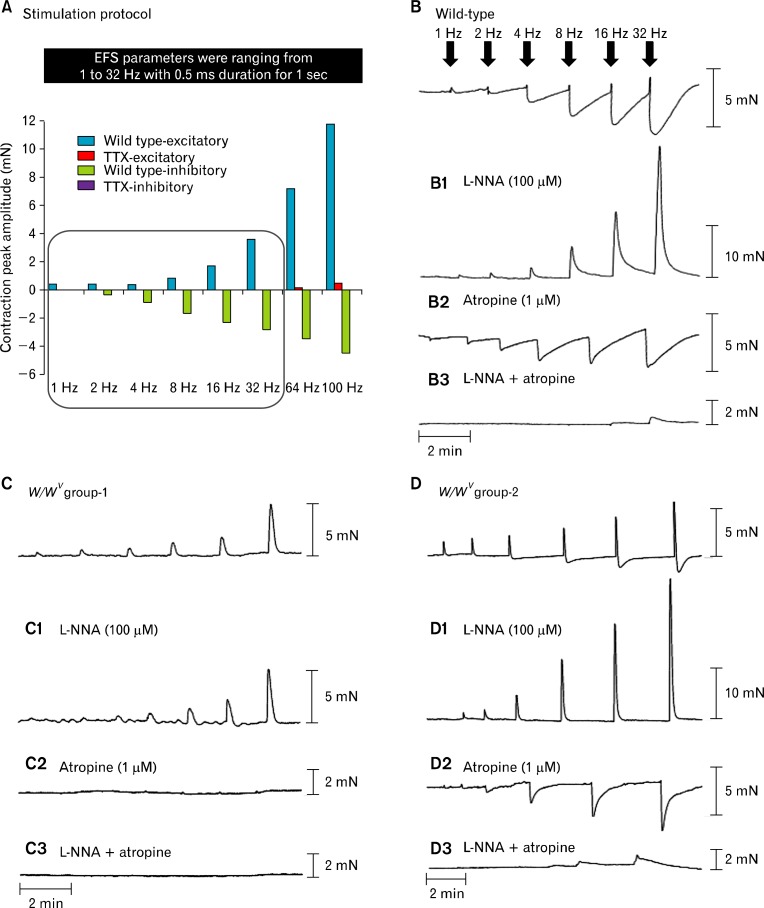
Figure 2.
Mechanical responses elicited by electrical field stimulation (EFS) in wild-type and W/WV gastric fundus. (A) Excitatory (blue) and inhibitory (green) responses to EFS (1–32 Hz; 0.5-millisecond duration for 1 second) were inhibited by tetrodotoxin (TTX, 1 μM). A small excitatory component persisted in the presence of TTX at 64 Hz and 100 Hz (red), and therefore we did not include these frequencies in subsequent analyses. (B) Representative traces of the mechanical responses in wild-type fundus induced by EFS under control conditions (1–32 Hz). (B1) Typical trace in the presence of L-NNA (100 μM). (B2) Responses after atropine (1 μM) and (B3) responses after the addition of Nω-nitro-l-arginine (L-NNA) and atropine. (C) shows responses from W/WV group 1 under control conditions and typical responses after (C1) L-NNA (100 μM), (C2) atropine (1 μM) and (C3) L-NNA and atropine. (D) Shows typical responses in W/WV group 2 under control conditions (D1) L-NNA (100 μM), (D2) atropine (1 μM) and (D3) L-NNA and atropine. Note the marked differences in mechanical responses to EFS between W/WV group 1 and W/WV group 2.

