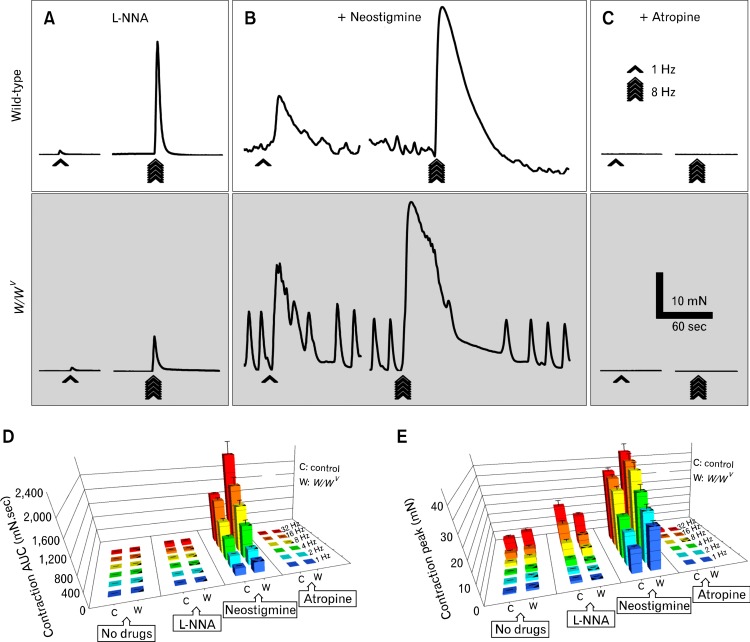
Figure 6.
Metabolic degradation of acetylcholine in W/WV fundus. (A) Representative traces of the mechanical responses in wild-type (white box) and W/WV (gray box) fundus induced by electrical field stimulation (EFS) in the presence of Nω-nitro-l-arginine (L-NNA, 100 μM), L-NNA and the cholinesterase inhibitor, neostigmine (3 μM) (B) and L-NNA, neostigmine and atropine (1 μM) together (C). Bar graphs represent the area under the curve (AUC) values (D) and peak amplitudes (E) of the excitatory responses to EFS in wild-type control mice (C) and W/WV (W) fundus, respectively. There were significantly greater contractions in W/WV muscles compared to wild-type. Note in the bar graphs the 1 triangle indicate a P-value < 0.05, 2 triangles indicate a P-value < 0.01 and 3 triangles indicate a P-value < 0.001.

