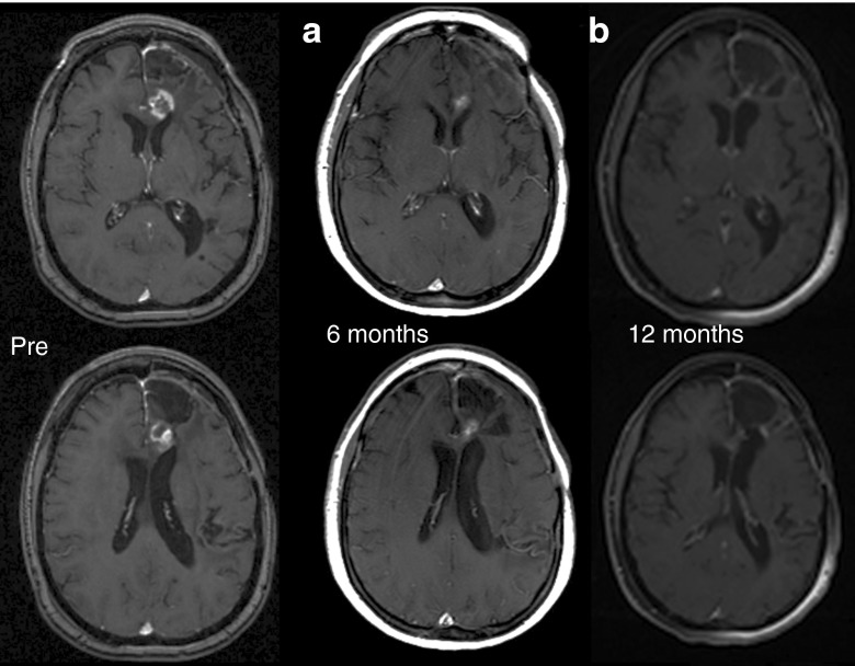
Figure 2.
Pre, 6- and 12-month posttreatment MRI scans. Shown are contrast-enhanced MRI scans from patient 13 with a recurrent glioblastoma multiforme who underwent a 72-hour infusion with 1 × 10¹⁰ TCID50 reovirus (highest dose tested). (a) MRI conducted at 6 months postinfusion, at formal end of trial, shows partial response compared to pre-entry MRI. (b) MRI conducted at 12 months postinfusion, on routine follow-up, and without any further antineoplastic therapy, shows a complete response compared to pre-entry MRI. MRI, magnetic resonance imaging; TCID50, tissue culture infectious dose50

