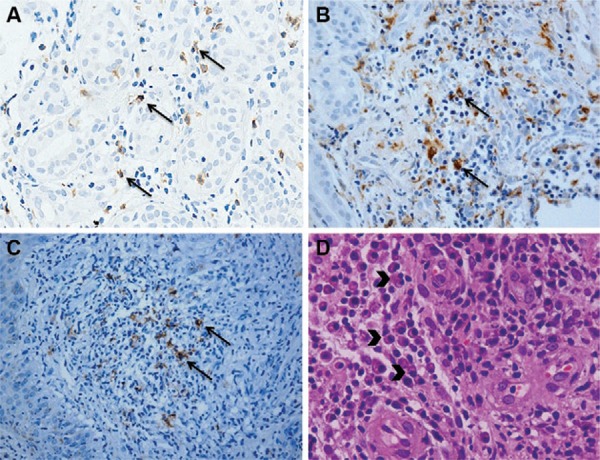
Fig. 2A. : immunostained CD4+ T cells (400X) (arrows); B: immunostained CD68+ cells (400X) (arrows); C: immunostained CD20+ cells (400X) (arrows); D: in haematoxylin and eosin, the presence of plasma cells in the inflammatory infiltrate (arrowheads) (400X).