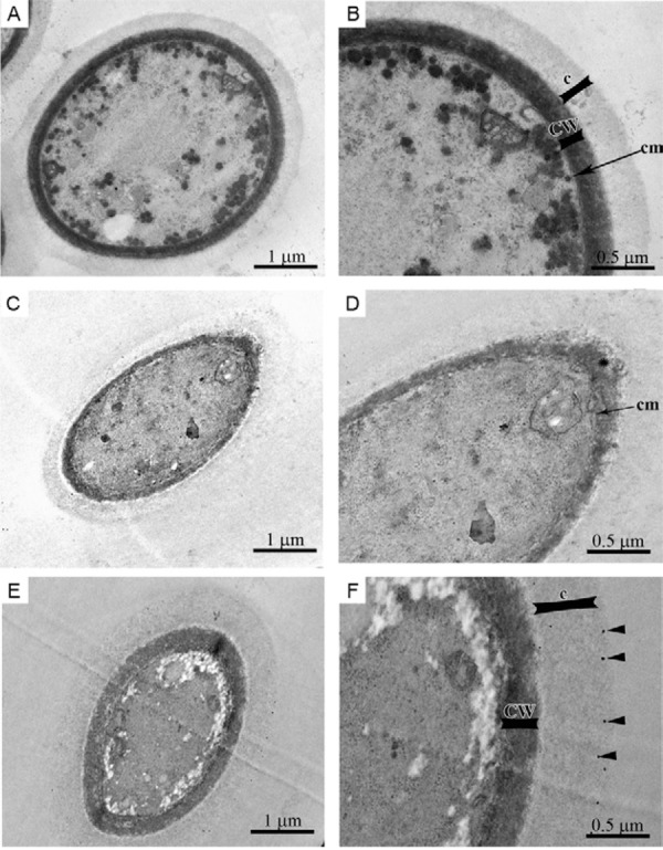
Fig. 5. morphological alterations of Cryptococcus neoformans treated with sub-inhibitory concentrations of silver nanoparticles (SNPs) (0.21 µg/mL) for 72 h at 35ºC. The untreated yeast exhibit a compact cell wall (CW), continuous cytoplasmic membrane (cm), homogeneous and electron-dense cytoplasm and a polysaccharide capsule (c) surrounding the cell (A, B). By contrast, yeasts treated with SNPs had a disrupted cytoplasmic membrane and CW (asterisk) and increased cell wall thickness (CW) (C, D). The SNPs appear to be retained in the polysaccharide capsule (F, black arrowhead).