. 2014 Mar 7;109(2):210–219. doi: 10.1590/0074-0276140266

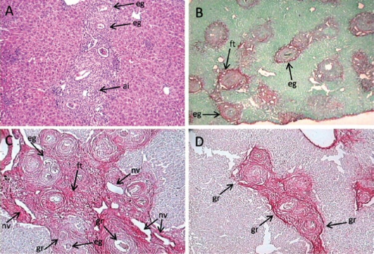
Fig. 2. : liver histopathology. A: well-nourished infected mouse with Schistosoma mansoni. Conglomerates of well-developed periovular granulomas around mature eggs, acute inflammatory cellular infiltration and scarce collagen deposition (60 days of infection) (haematoxylin-eosin 50X); B: undernourished-infected mouse with S. mansoni. Small scattered periovular granulomas with a poor infiltrate of polymorphonuclear cells around remnants of eggs (60 days of infection x 100, Picrosirius-red); C: well-nourished mouse. Periportal liver fibrosis (“pipestem”-like fibrosis) showing concentration of periovular granulomas, fibrous enlargement, increased vascularisation and chronic inflammatory infiltration of the portal space (150 days of infection, x 100, Picrosirius-red); D: undernourished mice. Groups of small periovular granulomas around empty eggshells, but absence of “pipestem”-like fibrosis (150 days of infection, x 100, Picrosirius-red); ai: acute inflammatory infiltrate; eg: eggshells (remnants); ft: fibrous tissue; gr: circumoval granulomas; nv: neovascularisation.

Fig. 2