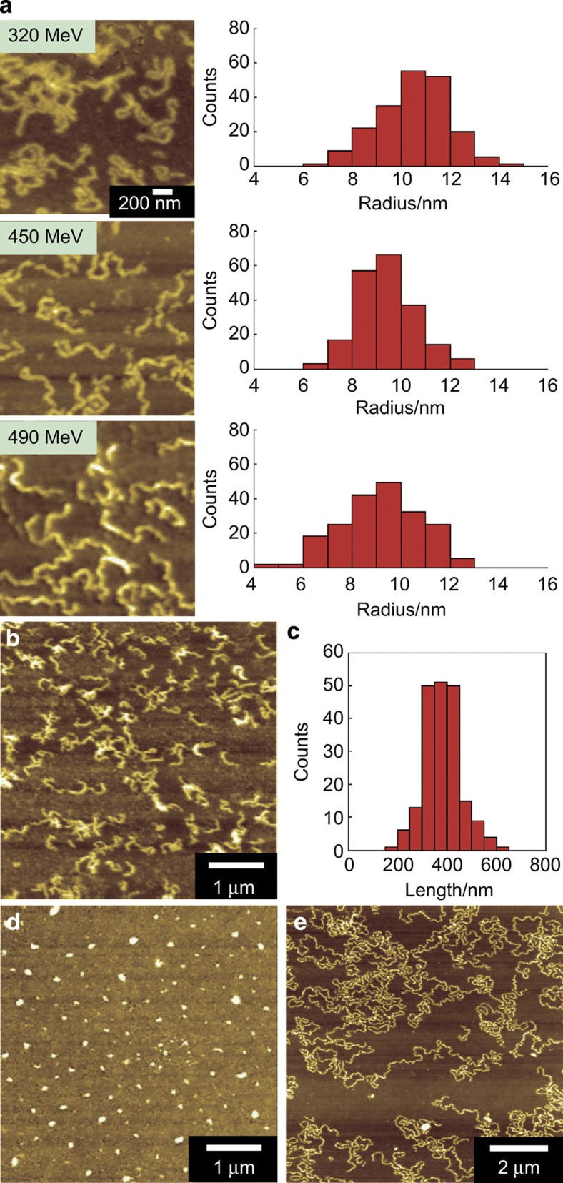
Figure 2. Effects of the type of charged particle and the film thickness.
(a) AFM images and radius distributions of HSA nanowires formed by irradiation of an HSA spin-coated film with various ion beams. The irradiation ion beams were 320 MeV 102Ru18+, 450 MeV 129Xe23+ and 490 MeV 192Os30+. (b) AFM image and (c) length distribution of the HSA nanowires formed by irradiation of a 500-nm-thick HSA spin-coated film with a 490 MeV 192Os30+-ion beam at a fluence of 1.0 × 10⁹ ions cm−2. AFM images of (d) HSA nanodots and (e) high aspect ratio HSA nanowires.

