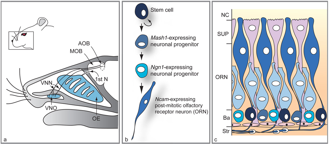
Figure 1.
Location of main olfactory epithelium (OE) and vomeronasal organ (VNO) in mouse head, and lineage and distribution of different cell types within the OE. (a) Sagittal section through an adult head reveals the position of the VNO relative to the OE. While the vomeronasal nerves (VNN) target the accessory olfactory bulb (AOB), axons of olfactory receptor neurons (first cranial nerve; 1st N) target the main olfactory bulb (MOB). (b) Scheme of the neuronal differentiation pathway in the OE. (c) Histological arrangement of cells in mature OE. Neuronal stem cells (black) located at the basal layer (Ba) adjacent to the stroma (Str) give rise to transit-amplifying progenitors expressing the Mash1 gene (round gray-blue cells) followed by Ngn1-expressing precursors (turquoise cells). The immature ORNs (light blue elongated cells) arise from the Ngn1-positive cells and mature into Ncam-expressing neurons (dark blue elongated cells). Supporting cells (SUP) lie in a single layer on the apical surface of the OE, at the nasal cavity (NC), while olfactory ensheathing cells wrap around nerve bundles in the underlying stroma. The VNO (not shown) has a similar histological arrangement, with basal cells at the boundary between epithelium and mesenchyme and neurons populating the bulk of the epithelium. Adapted from Kawauchi S, Beites CL, Crocker CE, et al. (2004) Molecular signals regulating proliferation of stem and progenitor cells in mouse olfactory epithelium. Developmental Neuroscience 26: 166–180, with permission. Copyright 2004 by S. Karger AG, Basel, Switzerland.

