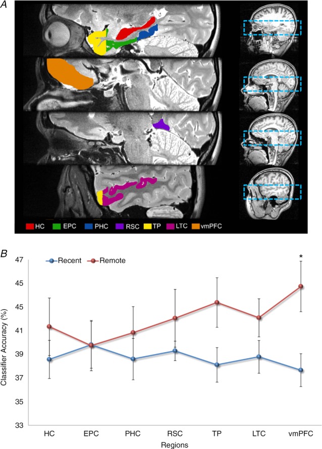
Figure 4.

A, the brain areas examined by Bonnici et al. (2012). The right panels show the bounding box of the high-resolution partial volume that was acquired for every subject. The left panels show the regions of interest that were demarcated, namely: hippocampus (HC), entorhinal and perirhinal cortices (EPC; combined because their responses were so similar), parahippocampal cortex (PHC), retrosplenial cortex (RSC), temporal pole (TP), lateral temporal cortex (LTC) and ventromedial prefrontal cortex (vmPFC). B, the MVPA results for memory decoding in each of the demarcated brain regions for recently formed autobiographical memories (blue) and for autobiographical memories that were formed 10 years ago (red). There was no significant difference in the classifier accuracy values for recent and remote memories in the hippocampus, but in vmPFC there was more accurate decoding of remote memories compared with recent memories (data from Bonnici et al. 2012; *P < 0.05; chance is 33%).
