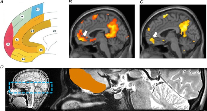
Figure 7.

A, a map of the human medial frontal cortex from Petrides & Pandya (1999; reproduced with permission from John Wiley & Sons). Examples of two conventional fMRI studies showing activity associated with autobiographical memory retrieval from Hassabis et al. (2007a2007a) in B and from Summerfield et al. (2009) in C. White arrows indicate the location of the activation in vmPFC that includes part of area 14, ventral parts of 24 and 32, the caudal part of area 10, and also some involvement of area 25. D, the vmPFC region analysed in the MVPA study of Bonnici et al. (2012) included these areas and more, specifically all of areas 14 and 25, ventral parts of areas 10, 24 and 32, and medial parts of area 11. The left panel shows the bounding box within which data were acquired on a T1-weighted structural MRI brain scan, and the right panel shows a close-up of an example T2-weighted structural MRI brain scan with the vmPFC region of interest delineated in orange.
