Figure 6.
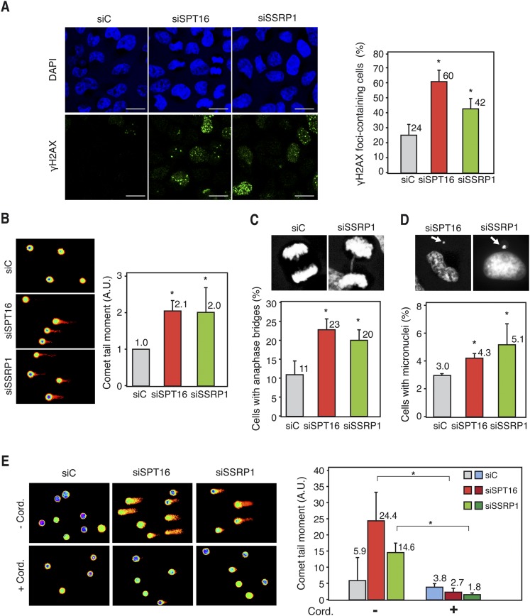
Genome instability in FACT-depleted human cells. (A) Immunofluorescence of γH2AX in siC (control), siSPT16, and siSSRP1 transfected HeLa cells. Nuclei were stained with DAPI. Graphics show the quantification of the percentage of cells containing γH2AX foci. Bars, 25 μm. More than 300 cells were analyzed in each case. (*) P < 0.05 (Student's t-test). (B) DNA breaks measured by single-cell gel electrophoresis (comet assay) of siC, siSPT16, and siSSRP1 transfected MRC-5 cells. Graphics show the median comet tail moment. More than 300 cells were analyzed in each case. (*) P < 0.05 (Mann-Whitney U-test). (C) Anaphase bridges in siC, siSPT16, and siSSRP1 transfected HeLa cells. Pictures show DAPI staining of control and SSRP1-depleted cells in anaphase. More than 100 anaphases were analyzed in each case. Other details are as in A. (D) Percentage of HeLa cells with microuclei after siC, siSPT16, or siSSRP1 transfection. Pictures show DAPI staining of SPT16- and SSRP1-depleted cells containing micronuclei. Other details are as in B. (E) Comet assay of siC-, SPT16-, or SSRP1-depleted HeLa cells after treatment or untreated for 4 h with 50 μM cordycepin. Other details are as in B. Mean and SD of three different experiments are shown.

