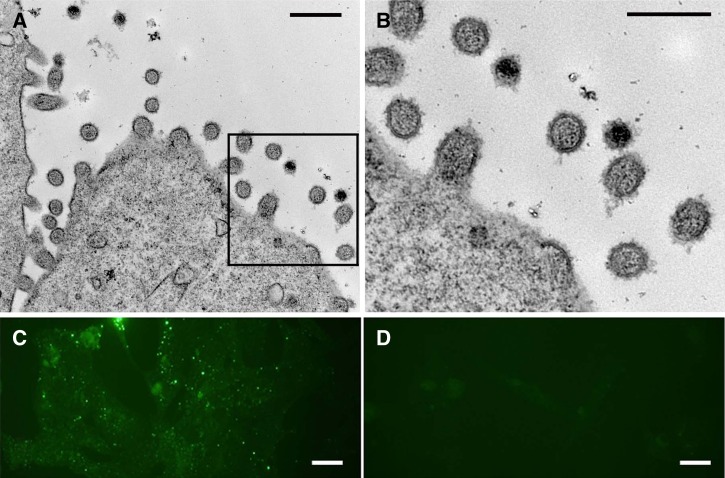
Figure 3.
(A and B) Electron micrographs showing classic hantavirus particles ranging from 80 to 120 nm in diameter budding from the surface of Vero cells. (C) Immunofluorescence assay showing characteristic punctuate, cytoplasmic staining of infected Vero E6 cells stained with anti-Seoul virus nucleocapsid antibody. (D) Mock-infected Vero E6 cells. Scale bars: A and B, 250 nm; C and D, 20 μm.

