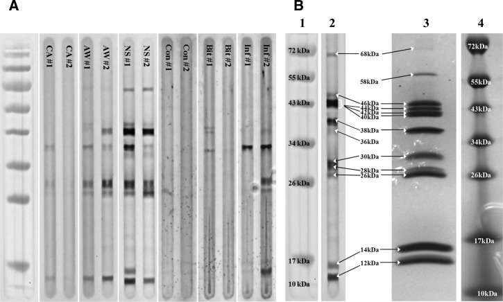
Figure 1.
Characterization of whole P. papatasi salivary gland protein extract sonicates and human host antibody reactions. (A) Immunoblot examples of plasma donors from three regions in Egypt and differentially exposed US military personnel with antibody specificity to P. papatasi salivary proteins. Lanes are designated by regional plasma donor codes (CA, AW, and NS) or differential exposure codes (Con = Control, Bit = Bitten, and Inf = Infected). (B) Comparison of immunoblot example and whole P. papatasi salivary gland protein sonicate. Lane 2 displays an immunoblot image of a North Sinai plasma donor with broad antibody specificities to P. papatasi salivary gland proteins. Lane 3 displays a Coomassie Brilliant Blue R-250-stained P. papatasi whole-salivary gland protein sonicate run on a denaturing SDS-PAGE gel. Lanes 1 and 4 are PageRuler protein MW ladders (Fermentas).

