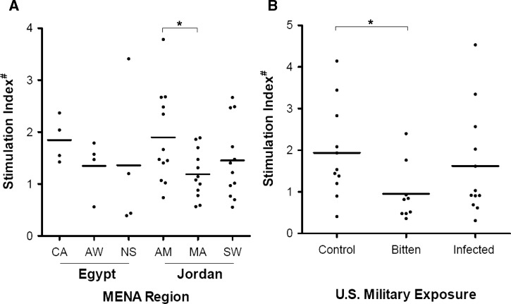
Figure 5.
Mixed leukocyte proliferation in vitro of peripheral blood donors after stimulation of MDDCs with P. papatasi SGS. (A) Representative sampling of regional Egyptian and Jordanian resident donor cellular proliferations when exposed to SGS-stimulated autogeneic MDDCs. Proliferation was measured using coculture with tritiated thymidine for 24 hours and β-particle decay counts per minute. Values presented are ratios of stimulated over unstimulated assays normalized to a single positive control donor proliferation run concurrently. (B) Proliferation data for US military personnel differentially exposed to sand fly bites and Leishmania parasites. *Significant differences by Student's t test (P < 0.05). #Stimulation index is the ratio of β-counts (3H-thymidine uptake) between unstimulated and SGS pre-stimulated MDDCs and mixed leukocyte cocultures.

