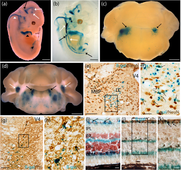
Figure 3.
Human MAOA-lacZ expressed in tyrosine hydroxylase (TH)-positive neurons in the locus coeruleus in adult brain, horizontal and ganglion cells in the adult retina, and several regions of the developing hindbrain. Expression analysis of the human MAOA-lacZ strain was examined using β-galactosidase (β-gal) histochemistry (blue). (a) E12.5 whole embryos stained in the prepontine hindbrain (black arrow) that extended to the basal midbrain and the prosomeres 1 and 2 of the diencephalon (white arrows). Staining extended from the pontine hindbrain (pons proper) to the medullary hindbrain (medulla) (white arrowhead). Staining was notable in the anterior part of the developing limbs (black arrowhead). (b) E12.5 cleared embryos additionally demonstrated staining in fibers of the prepontine hindbrain, and the pontine hindbrain. Staining was present in fibers of the medullary hindbrain that extended into the thoracic cavity; and fibers of the ventral region of the somites that started in the upper thoracic cavity, and extended towards the posterior limbs. (c) P7 brains showed staining in nuclei surrounding the fourth ventricle, a region where the locus coeruleus (LC) is located (black arrows). (d) Adult brains stained in the LC (black arrows), and lateral cerebellar nuclei (white arrows). Staining was also present in the medial vestibular nuclei, and lateral vestibular nuclei. (e) Colocalization experiment using β-gal staining and neuronal nuclei (NeuN) immunohistochemistry (brown) performed on adult brain cryosections suggested expression of MAOA-lacZ in mature neurons in the LC. Sparse staining was detected in neurons populating the medial parabrachial nucleus. Boxed region in (e) is shown in (f). (f) Higher magnification revealed expression of β-gal in neurons in the LC (black arrows). (g) Colocalization experiment using β-gal staining and an anti-TH immunohistochemistry (brown) performed on adult brain cryosections suggested expression of MAOA-lacZ in TH-positive neurons populating the LC. Boxed region in (g) is shown in (h). (h) Higher magnification revealed expression of β-gal in TH-positive neurons in the LC (black arrows). (i) Staining was detected in the retina, extending from the inner limiting membrane (black arrows) to the outer plexiform layer (white arrows), and the outer limiting membrane (OLM). (j,k) Colocalization experiment using β-gal staining and an anti-calbindin (anti-Calb) immunohistochemistry (brown) performed on adult eye cryosections suggested expression of MAOA-lacZ in horizontal cells populating the OPL (white arrows), and ganglion cells populating the ganglion cell layer (black arrows). Boxed region in (j) is shown magnified in (k). INL, inner nuclear layer; IPL, inner plexiform layer; MBP, medial parabrachial nucleus; ONL, outer nuclear layer; V4, fourth ventricle. Scale bar: (a-d) 1 mm; (e,g) 100 μm; (f,h) 25 μm; (i-k) 20 μm.

