Figure 5.
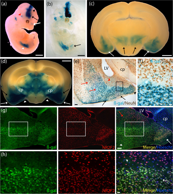
Human NR2F2-lacZ expressed in mature neurons populating the basolateral and corticolateral amygdaloid nuclei that are immunoreactive for the Nr2f2 mouse protein. Expression analysis of the human NR2F2-lacZ strain was undertaken by examination of β-galactosidase (β-gal) staining (blue). (a) E12.5 whole embryos revealed staining in the rostral secondary prosencephalon (black arrow) that extended throughout all three prosomeric regions of the diencephalon (black arrowhead). Staining was present in the nasal cavity (white arrow), the vestibulochochlear ganglion (red arrowhead) and mesenchyme of the posterior limbs (white arrowhead). (b) E12.5 cleared embryos additionally demonstrated staining in the developing bladder (black arrow). (c) P7 brains stained in the amygdala nuclei (white arrows), and the subthalamic nuclei (black arrows). (d) Adult brains revealed strong staining extending from the posterior basolateral amygdaloid nuclei (BLP) (black arrows) to the posterolateral cortical amygdaloid nuclei (PLCo) (white arrows), and the posteroventral part of the medial amygdaloid nuclei (MePV) (white arrowheads). Broad staining was detected in the ventral thalamic area, excluding the cerebral peduncle (cp). (e) Colocalization experiment using β-gal staining and a neuronal nuclei (NeuN) antibody (brown) performed on adult brain cryosections revealed strong expression of NR2F2-lacZ in mature neurons populating the BLP, and the basomedial amygdaloid nuclei (BMP) (red arrows). Colocalization was found in the PLCo, and the posteromedial cortical amygdaloid nuclei (PMCo) (black arrows), and the MePV (black arrowhead). Lower level of β-gal staining was detected in mature neurons in the anterolateral amygdalohippocampal area (AHiAL) (red arrowhead). Boxed region in (e) is shown in (f). (f) Higher magnification revealed strong expression of β-gal in mature neurons in the PMCo and sparse expression in mature neurons in the AHiAL. (g) Colocalization experiment, using an anti-β-gal antibody (green), and an NR2F2 antibody (red), performed on adult brain cryosections revealed strong β-gal labeling in cells expressing the Nr2f2 mouse gene in brain regions extending from the PMCo (white arrowhead) to the MePV (white arrow). Lower levels of β-gal were detected in the AHiAL (red arrow). Boxed region in (g) is shown in (h). (h) Higher magnification revealed strong expression of β-gal (green) in Nr2f2-positive cells (red) in the PMCo (white arrow) and lower expression in the AHiAL (white arrowhead). LV, lateral ventricle. Scale bar: (a-d) 1 mm; (e,g) 100 μm; (f,h) 20 μm.

