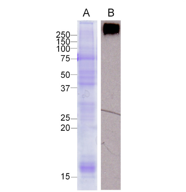
Figure 1.

Western blot analysis of hemocytin in hemocytes of Bombyx mori. Samples of normal hemocytes were separated by 12.5% SDS-PAGE and stained by Coomassie Brilliant Blue (A). The western blot analysis was conducted (B) as described in the Materials and Methods section. High quality figures are available online.
