
Figure 3.
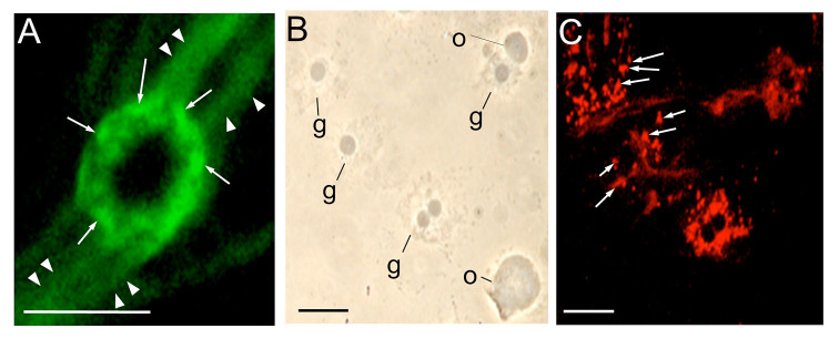
Immunostaining of the accumulated hemocytin in the granules of granulocytes of Bombyx mori. After collection in a microfuge tube with a solution of 1 M DTT, hemolymph was smeared on glass slides, fixed, stained with anti-hemocytin antiserum in combination with secondary antiserum labeled with Alexa Fluor 488 (A) or Alexa Flour 594 (C) and observed under fluorescence microscopy (A, C) or phase contrast microscopy (B). The scale bars indicate 10 µm. The arrows point to granules in the granulocyte. The arrowheads indicate fibrous structures. g, granulocyte; o, oenocytoid. High quality figures are available online.