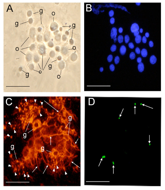
Figure 5.

Immunostaining of hemocytin in a small aggregate made in vivo after Escherichia coli injection. Thirty seconds after fluorescein pre-stained E. coli injection, the hemolymph of Bombyx mori larvae was placed on glass slides, smeared, fixed, and stained with DAPI (B) or anti-hemocytin antiserum in combination with Alexa Fluor 594-labeled secondary antiserum (C). (A) A phase contrast microscopy image of an aggregate of hemocytes is shown. (B) A DAPI-stained image of (A) indicating hemocyte nuclei is shown. (C) An Alexa Fluor 594-stained image of (A) indicating hemocytin is shown. (D) A fluorescein-stained image of (A) indicating pre-stained E. coli cells is shown. The scale bars indicate 20 µm. The arrows show fibrous structures around hemocytes. The arrowheads indicate granules spreading from burst granulocytes. g, granulocyte; o, oenocytoid. High quality figures are available online.
