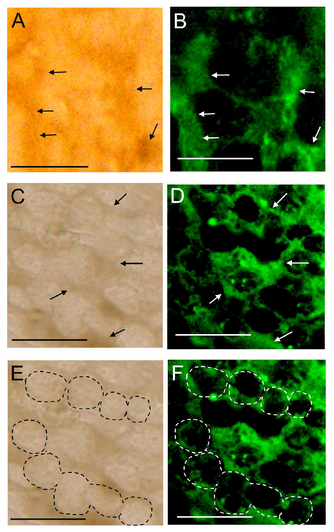
Figure 7.

Immunostaining by the anti-hemocytin antiserum of the nodule formed in Bombyx mori fifth-instar larvae 10 min after injection with Escherichia coli cells and 30 min after injection with Micrococcus luteus. Paraffin sections of the nodules induced by either E. coli (A and B) or M. luteus (C–F) injection were stained with anti-hemocytin antiserum in combination with Alexa Fluor 488-labeled secondary antiserum and observed under bright field (A, C, and E) and fluorescence (B, D, and F) microscopy. Under bright field microscopy, the granulocyte cell bodies were observed as bright regions, as indicated by dashed circles in (E), and the matrix was dark, as indicated by the arrows in (A and C). The scale bars indicate 10 µm. High quality figures are available online.
