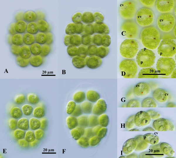
Figure 1.
Light microscopy of vegetative colonies of two species of Colemanosphaera. (A)-(D) C. charkowiensis (Korshikov) Nozaki et al. comb. nov. 2013-0615-IC-7. (A), (B) Two views of a 32-celled colony shown at the same magnification. (A) Surface view. (B) Optical section. (C), (D) Two views of cells shown at the same magnification. (C) Surface view showing contractile vacuoles (cv) distributed in only the anterior end of cells. (D) Optical section. Note multiple pyrenoids (p) and strong longitudinal striations in the chloroplast periphery. (E)-(I) C. angeleri Nozaki sp. nov. 2010-0126-1. (E), (F) Two views of a 32-celled colony shown at the same magnification. (E) Surface view. (F) Optical section. (G)-(I) Three views of cells showing contractile vacuoles (cv) and pyrenoids (p), shown at the same magnification throughout.

