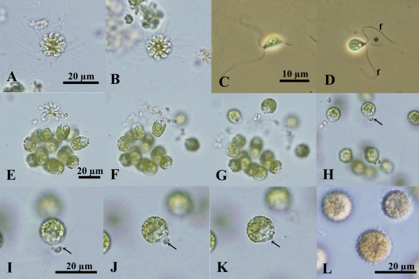
Figure 3.

Light microscopy of sexual reproduction in Colemanosphaera charkowiensis (Korshikov) Nozaki et al. comb. nov. (A), (B), (E)-(L) 2013-0615-IC-3x4x7. (C), (D) 2010-0713-E5. (A), (B) Sperm packets (bundles of male gametes) shown at the same magnification. (C), (D) Male gametes shown at the same magnification. Note cytoplasmic protrusion (asterisk) near the base of the flagella (f). (E)-(K) Successive stages of male and female gamete release and conjugation. Note male gamete (arrow) fusing with female gamete. (E)-(H) Shown at the same magnification throughout. (I)-(K) Shown at the same magnification throughout. (L) Mature aplanozygotes.
