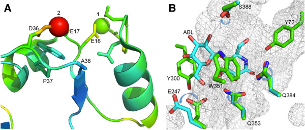
Figure 5.

Structural analysis of GOOX-T1. (A): Metal binding sites of the FAD-binding domain (coloring by b-factor); A38 is close to the zinc ion 2 bound to D36 and E17. (B): Residues for mutation in relation to the substrate analog, 5-amino-5-deoxy-cellobiono-1,5-lactam (ABL); the movement of residues in the absence of ABL (PDB ID: 1ZR6, green) compared with the presence of ABL (PDB ID: 2AXR, cyan) are indicated by arrows.
